Thursday 30 June 2016

Myocardial crypts in hypertrophic cardiomyopathy

Over recent years there has been interest, and debate, about the significance of myocardial crypts seen on cardiac MRI scanning. Crypts (also known as clefts, crevices or fissures) are defined as an invagination of the myocardium that penetrates >50% of the myocardial thickness in diastole. An example is shown in the figure below: here there are two crypts in the inferior wall.

Two inferior wall myocardial crypts (arrowed)

To see this image as a cine CMR video, click on the video below to play it:


So are myocardial crypts significant? It has been suggested that they may represent a 'prephenotypic marker' of hypertrophic cardiomyopathy, based upon a 2012 paper in which crypts were  found with a high prevalence in genotype-positive (phenotype-negative) hypertrophic cardiomyopathy patients, but in none of a normal control group.

More recently, however, a study has shown that they are not uncommonly seen as an incidental finding in cardiac MRI studies, with an overall prevalence of 6.7%, although the prevalence is higher in patients with hypertrophic cardiomyopathy, myocarditis and hypertension.

So are myocardial crypts significant or not? The jury is still out, but for now the incidental finding of myocardial crypts, especially if multiple, should always prompt a careful review for any other imaging findings, clinical features or family history that might point towards a diagnosis of hypertrophic cardiomyopathy.


Monday 27 June 2016

Hypokinetic non-dilated cardiomyopathy: a new category of cardiomyopathy

The European Society of Cardiology working group on myocardial and pericardial diseases has recently published a position statement on the diagnosis of dilated cardiomyopathy, and in doing so they have proposed a new diagnostic category: hypokinetic non-dilated cardiomyopathy, or HNDC.

CMR showing an enlarged left ventricle in dilated cardiomyopathy

How is this new category defined? The ESC working group proposes that:

Hypokinetic non-dilated cardiomyopathy is "left ventricular or biventricular global systolic dysfunction without dilatation (defined as LVEF <45%), not explained by abnormal loading conditions or coronary artery disease"

The term 'abnormal leading conditions' refers to cases where there is hypertension or valvular heart disease that has placed abnormal pressure and/or volume loading on the left ventricle, leading to dysfunction.

So why create this new category of HNDC? The reason is that, put simply, dilated cardiomyopathy doesn't always involve left ventricular dilatation. It's been recognised that in some cases there is clear evidence of left ventricular systolic dysfunction, but in the absence of left ventricular dilatation. For example, this has been described in carriers of Lamin A/C gene mutations. To avoid confusion about diagnostic terminology, this new category of HNDC has been proposed.

The new document is well worth a read. As well as the new category of HNDC, it also provides useful guidance on the investigation of dilated cardiomyopathy, and on diagnostic criteria in relatives.

If you'd like to read the guidelines, they can be accessed here:

Thursday 23 June 2016

The prognostic value of TAPSE in acute PE

Bedside echocardiography has a useful role to play in the identification of right ventricular dysfunction in acute pulmonary embolism. But what's the best way to identify right ventricular dysfunction?

One commonly used criterion is the ratio between right ventricular and left ventricular diameters, measured in a 4-chamber view (which can be done either on echo or on CT). An RV:LV ratio of >0.9 is used to define right ventricular dysfunction in this setting, and has been included in guidance by the American Heart Association.

However, a paper by Pruszczyk and colleagues advocates the use of TAPSE as a better option than RV:LV ratio.

TAPSE is tricuspid annulus plane systolic excursion, and is simply measured using M-mode echo in an apical 4-chamber view. The M-mode cursor is placed at the tricuspid annulus, and the longitudinal displacement of the annulus during systole is measured in millimetres (see illustration).

Measurement of TAPSE using M-mode echo


Pruszczyk and colleagues found that a TAPSE of 15mm or less was associated with a higher risk of 30-day mortality from acute pulmonary embolism, whereas a TAPSE of >20mm was associated with a very low risk of mortality. The authors found that TAPSE performed better than RV:LV ratio in 30-day risk stratification.

If you'd like to read their original paper, it can be accessed by clicking on the reference below:

Monday 20 June 2016

An incidental finding next to the heart on a CT scan

During cardiac imaging, it's not unusual to make unexpected findings outside the heart. Indeed, during cardiac MRI scanning incidental extracardiac findings are reported in around one-fifth of cases, and during coronary CT scanning the reported prevalence of incidental findings is even higher.

The image below shows a CT scan, and there's an incidental finding of a 'mass' adjacent to the right atrium. So what is the nature of this mass?

CT scan - the 'mass' next to the heart is arrowed

This is a pericardial cyst - an uncommon abnormality. On CT scanning pericardial cysts are well-defined, thin-walled and do not enhance with contrast. On cardiac MRI (see example below) fluid-filled pericardial cysts appear hypointense on T1-weighted images, and hyperintense on T2-weighted images, and do not enhance with gadolinium contrast.

Pericardial cyst on cardiac MRI

Most cases are congenital cysts, but they can also be acquired following cardiac surgery, trauma or pericarditis. Pericardial cysts are found in 1 per 100,000 individuals and can occur anywhere within the pericardium, although they are more commonly found adjacent to the right heart than the left.

Pericardial cysts are usually asymptomatic, but in some cases present with chest pain, palpitations, breathlessness or cough. They are usually benign, but (rarely) life threatening complications such as cardiac tamponade have been reported.

Pericardial cysts are usually managed conservatively, but percutaneous aspiration (sometimes with ethanol sclerosis) or surgical resection can be considered if the patient is symptomatic or in the event of life-threatening complications.

You can read more about pericardial cysts in the following review:


Information about the management of pericardial cysts can be found in the ESC 2015 Guidelines for the Diagnosis and Management of Pericardial Diseases.

Tuesday 14 June 2016

Ten key facts about coarctation of the aorta

Coarctation of the aorta is a narrowing of the aorta, classically (but not always) just distal to the origin of the left subclavian artery. Here are ten key facts about coarctation of the aorta to be aware of in your cardiology clinic.

MR aortogram showing coarctation of the aorta plus collaterals
  1. Coarctation of the aorta accounts for 5-8% of congenital heart defects, with a prevalence of around 0.03% of live births
  2. The location of coarctation of the aorta is most commonly at the insertion point of the ductus arteriosus (just distal to the origin of the left subclavian artery)
  3. Ectopic coarctation can sometimes occur in the ascending aorta, the descending aorta or even the abdominal aorta
  4. The region of coarctation is most commonly discrete, but it can sometimes extend over a long segment of the aorta. There is often some dilatation of the aorta immediately beyond the coarctation
  5. Bicuspid aortic valve is a frequent association with coarctation of the aorta, reported in up to 85% of cases. Other associations with coarctation of the aorta include Turner syndrome, aortic stenosis (subvalvular, valvular or supravalvular), Shone syndrome and neurofibromatosis
  6. Mild cases of coarctation of the aorta may not present until adulthood. Common presentations include incidental hypertension (affecting the upper body) or a murmur
  7. A blood pressure gradient >20mmHg between upper and lower limbs indicates significant coarctation of the aorta
  8. Chest X-ray findings include rib notching (caused by collaterals) and a double contour of the descending aorta ('figure 3 sign')
  9. Both MRI (see image above) and cardiac CT are useful non-invasive methods for imaging the anatomy of coarctation of the aorta, and the presence of collateral vessels
  10. Treatment options include stenting, resection with end-to-end anastomosis, and resection with interposition of a graft

After primary repair of coarctation of the aorta, long-term survival remains below that of normal controls, and the need for re-operation is relatively common. Patients should therefore remain under lifelong surveillance after surgical repair.

For further reading about the importance of ongoing surveillance after surgical repair, read the following reference:

Wednesday 8 June 2016

Ten key facts about ARVC

Arrhythmogenic right ventricular cardiomyopathy (ARVC, also known as arrhythmogenic right ventricular dysplasia or ARVD) is one of the genetic cardiomyopathies. Here are ten key facts about ARVC that you should be aware of in your cardiology clinic.

Cardiac MRI showing ARVC (4-chamber view)

  1. ARVC predominantly affects the right ventricle, causing the replacement of myocardium by fibrofatty tissue. However it can also involve the left ventricle as the disease progresses
  2. The population prevalence of ARVC is thought to be somewhere between 1 in 2,500 and 1 in 5,000
  3. ARVC has an autosomal dominant pattern of inheritance (with incomplete penetrance and variable expression), and affects males more often than females (ratio 2.7:1)
  4. The genetic mutations causing ARVC affect the desmosomal proteins, which connect cells and play a role in signal transduction
  5. Common presentations of ARVC include syncope and sudden cardiac death
  6. The diagnosis of ARVC depends upon the presence of major and/or minor criteria (see Task Force reference below)
  7. ECG criteria include the presence of T wave inversion in leads V1-V3 and epsilon waves
  8. Cardiac imaging (e.g. echo, CMR, contrast ventriculography) plays an important role in the diagnosis of ARVC but the diagnosis must not be based solely upon imaging findings, but instead must be based upon Task Force criteria
  9. Cardiac biopsy is of limited value in the diagnosis of ARVC due to the segmental nature of the disease
  10. Treatment options in ARVC include antiarrhythmic drugs, catheter ablation therapy and/or ICD implantation. Treatment with heart failure drugs may also be indicated

You can read more about ARVC in these key references:


Marcus FI, et al. Diagnosis of Arrhythmogenic Right Ventricular Cardiomyopathy/Dysplasia: Proposed Modification of the Task Force Criteria. Circulation 121: 1533-1541.

Monday 6 June 2016

How to assess myocardial viability on CMR

In last week's blog, I described how useful cardiac MRI is in the diagnosis of myocardial infarction. The presence of subendocardial late gadolinium enhancement is both a sensitive and specific indicator of the presence of an area of infarction. But what about myocardial viability? We're often asked to assess whether an infarcted region of myocardium has sufficient residual viability to justify coronary revascularization - but how do we do this?

Well, there are two ways: a quick way, and a slightly longer (but more accurate) way.


Viable versus non-viable myocardium on CMR

The quick way simply involves looking at the myocardium, and judging how much of the myocardial thickness is infarcted - i.e., what proportion of the myocardial thickness is occupied by late gadolinium enhancement (LGE). If the LGE occupies less than 50% of the thickness, then we judge the segment overall to still be viable. If the LGE occupies more than 50%, then it's no longer viable. If it's exactly 50%, then we say there's 'partial' viability.

This method is quick and easy, but it's difficult to be precise about the percentages, and it's hard to accept that the difference between 49% and 51% is so clear-cut (especially when it has big implications for revascularization decisions).

A more refined method is to say that if the LGE occupies <25% of the myocardial thickness then it's viable, and >75% means it's non-viable. If it's between 25-75%, then it's indeterminate and we need go on to perform a dobutamine stress CMR study to see whether the myocardial contractility improves with low-dose stress. If it does, then it's viable; if it doesn't, then it isn't.

If you'd like to learn more about the use of CMR in the assessment of myocardial infarction and viability, then check out my Cardiac MRI Essentials course at Medmastery.

Friday 3 June 2016

CMR and the diagnosis of myocardial infarction

How can we tell if a patient has had a previous myocardial infarction? I guess we could look for Q waves on their ECG, or for regional wall motion abnormalities on echocardiography. But one of the best ways is using CMR, which shows even small areas of infarcted myocardium with clarity. So how do we do this?

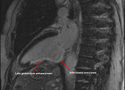
CMR: Inferior MI, with inferobasal aneurysm

The technique uses gadolinium, an intravenous contrast agent commonly given during MRI studies. In stress cardiac MRI, for instance, gadolinium is used to visualise myocardial perfusion in the assessment of coronary artery disease.

When gadolinium is given, it enters normal myocardium quickly and is then rapidly washed out by normal perfusion. If you scan a normal heart ten minutes after a bolus of gadolinium is given, there's normally no gadolinium left behind in the myocardium.

However if there is myocardial scar tissue present as a result of an infarction, the gadolinium does not get washed out so quickly. Instead, the gadolinium stays in the infarcted areas of myocardium much longer than normal, and is still visible if imaging is done ten minutes later.

So to identify areas of infarction, we give a bolus of gadolinium IV, and then we wait for ten minutes. We then scan the heart to look for 'bright' areas of infarcted myocardium, which indicates that gadolinium is still present. We call this 'late gadolinium enhancement', or LGE.

An example is shown above. This patient has had an inferior myocardial infarction in the past, caused by occlusion of their right coronary artery. We can see evidence of LGE in the inferior wall of the left ventricle, corresponding to the area of infarction. You'll also notice a bulge in the basal inferior wall, indicating an inferobasal aneurysm.

You'll see that the LGE is subendocardial rather than epicardial. This is because myocardium infarcts 'from the inside to the outside'. In other words, the subendocardium is always involved in an infarction, but the extent to which the infarction extends outwards varies from case to case. In some cases the LGE stays subendocardial, in others it can be 'full thickness'.

The extent of LGE also tells us about residual myocardial viability, but that's a topic for another blog...

If you'd like to learn more about the use of LGE in the CMR diagnosis of myocardial infarction, then check out my Cardiac MRI Essentials course at Medmastery.